- Shanghai, China
- [email protected]
- +86-21-58189111
The development history, basic structure and common complications of arthroscopy.
Arthroscopy originated in Japan at the beginning of the 20th century, and developed rapidly in the United States and other countries after the 1970s. Over the past few decades, arthroscopy has revolutionized the diagnosis and treatment of intra-articular disease. Arthroscopy can provide a comprehensive observation of intra-articular structures, which is more subtle than incision surgery, and many intra-articular structures and lesions can be directly observed and treated. Some people call arthroscopic technology, internal fracture fixation and artificial joint replacement as the three major advances in the field of orthopedics in the 20th century. Arthroscopy has become widely accepted and used to be called a "young man's toy" and is now the standard diagnostic and therapeutic technique. Arthroscopy is an important part of joint surgery, which fully reflects the development trend of minimally invasive modern surgery.
With the gradual popularization of endoscopic technology in my country, arthroscopic surgery has become a routine diagnosis and treatment method in bone and joint surgery. At present, among the various system endoscopes of the whole body, arthroscopy is the most comprehensive and widely used, and thus forms an independent arthroscopic surgery. The arthroscopic surgery business has become a growth point for hospital and department performance.
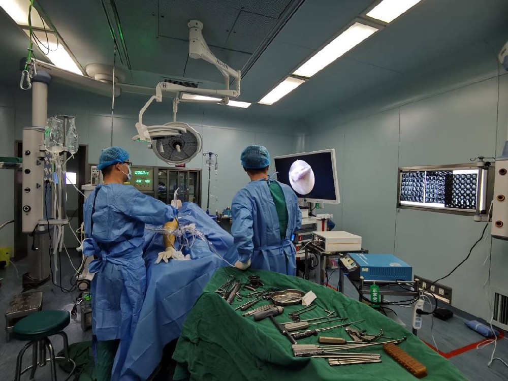
The basic structure of the arthroscope is an optical system, the center is the rod lens system for image acquisition, the surrounding is the optical fiber leading into the light source, and the outside is the metal protective sheath. By creating a small incision of about 0.8mm~1.0cm in the skin, placing the arthroscope into the joint, and connecting the camera and display equipment behind it, the shape and lesions in the joint can be directly observed, and by using special instruments, the joint can be inspected. Internal disease can be treated, thus avoiding many arthrotomies.
The basic structure of the arthroscope is an optical system, the center is the rod lens system for image acquisition, the surrounding is the optical fiber leading into the light source, and the outside is the metal protective sheath. It consists of arthroscopic lens, camera, host, display and cold light source. The arthroscopic lens is a thin rod with a length of more than 20 cm and a thickness of 4-5 mm, which is used to insert into the joint cavity. The rod contains a set of optical fibers and a set of fluoroscopy. Image sent. Outside the joint, an optical cable connects the optical fiber to the cold light source, so that the cold light source can illuminate the joint; a camera connects the lens with the host and the monitor, so that the image inside the joint is reflected on the monitor. By creating a small incision of about 0.8mm~1.0cm in the skin, placing the arthroscope into the joint, and connecting the camera and display equipment behind it, the shape and lesions in the joint can be directly observed, and by using special instruments, the joint can be inspected. Internal disease can be treated, thus avoiding many arthrotomies.

Complications of arthroscopic knee surgery: postoperative infection, neurovascular injury behind the knee joint, joint adhesions, lower extremity deep vein thrombosis, etc.
Complications of shoulder arthroscopy: tracheal compression and upper airway obstruction caused by extravasation of a large amount of joint irrigation fluid; cerebral ischemic injury; arrhythmia caused by abnormal absorption of epinephrine in the irrigation fluid into the blood; tension pneumothorax; pulmonary Air embolism, etc.
Complications of arthroscopic elbow surgery: hematoma, delayed wound healing, persistent exudate, superficial infection, transient nerve injury, deep infection, persistent nerve injury without recovery, etc.
Leave a Comments