Laparoscopic resection of hilar cholangiocarcinoma, also known as laparoscopic hepatopancreatoduodenectomy (HPD), is a minimally invasive surgical technique used to remove tumors located at the junction of the bile ducts and liver. This procedure is complex and requires expertise in both laparoscopic and hepatobiliary surgery.
Preoperative evaluation:
Before the operation, a comprehensive evaluation of the patient is necessary. This includes a detailed medical history, physical examination, blood tests, imaging studies such as MRI and CT scans, and endoscopic retrograde cholangiopancreatography (ERCP) to assess the extent of the tumor and its relationship to the bile ducts.
Surgical technique:
The procedure is performed under general anesthesia, and the patient is placed in a modified lithotomy position with the legs apart. A laparoscope is introduced through a small incision in the umbilicus, and additional ports are placed in the upper abdomen to accommodate other surgical instruments. Carbon dioxide gas is then insufflated into the abdomen to create a working space.
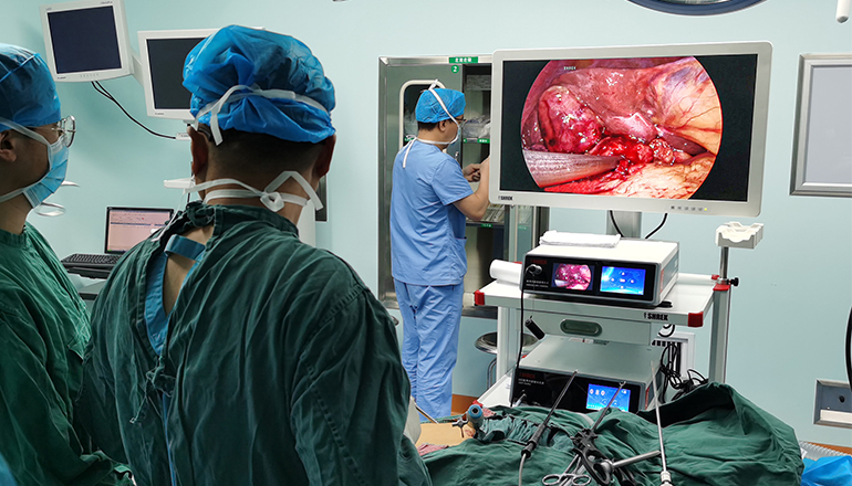
The first step is to mobilize the liver and expose the hepatic hilum. This involves dividing the ligamentum teres and falciform ligament, and retracting the left lobe of the liver to expose the common bile duct and hepatic artery. The lymph nodes around the hepatic hilum are dissected and removed for pathological evaluation.
Next, the porta hepatis is dissected to expose the right and left hepatic ducts, as well as the portal vein and hepatic artery. The right and left branches of the hepatic ducts are then dissected and divided using a stapler, and the specimen is removed through a small incision in the upper abdomen.
After the specimen is removed, the bile duct and pancreatic duct are reconstructed using a Roux-en-Y hepaticojejunostomy and pancreaticojejunostomy. The remaining ports are then removed, and the abdominal incisions are closed.
Postoperative management:
Patients are typically kept in the hospital for several days following surgery to monitor their recovery. Pain medication, antibiotics, and anti-nausea medications are administered as needed. Patients are allowed to resume eating and drinking once bowel function has returned. A follow-up appointment with the surgeon is scheduled within several weeks after the surgery.
Conclusion:
Laparoscopic resection of hilar cholangiocarcinoma is a complex and technically challenging procedure that requires expertise in both laparoscopic and hepatobiliary surgery. However, in experienced hands, this approach has been shown to be safe and effective, with a shorter hospital stay, less postoperative pain, and faster recovery compared to traditional open surgery.
Long-term outcomes of laparoscopic resection of hilar cholangiocarcinoma are still being evaluated, but early results suggest that it may have comparable oncological outcomes to open surgery. However, patient selection is crucial, and only carefully selected patients with early-stage tumors and no evidence of distant metastasis should be considered for this procedure.
In conclusion, laparoscopic resection of hilar cholangiocarcinoma is a complex and demanding procedure that requires advanced surgical skills and experience. However, it offers several benefits over open surgery, including shorter hospital stay, less postoperative pain, and faster recovery. Careful patient selection and meticulous surgical technique are essential to achieve optimal outcomes. Therefore, it is important to seek out an experienced surgical team who can provide the best possible care for patients with this challenging disease.
Leave a Comments