Foraminal endoscopic nucleectomy is a surgical method to remove the nucleus pulposus, nerve roots, diseased tissue and hyperplastic bone tissue in the dural sac through a foramenoscope.
Disease information
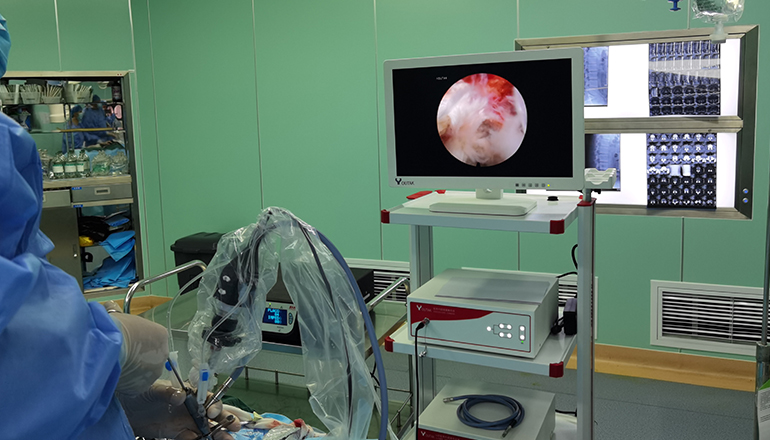
Similar to the spinal endoscope, the foramoscope is a tube equipped with light, which enters the intervertebral foramen from the side or the back of the patient's body (either flat or oblique), and performs surgery in the safe working triangle area. When surgery is performed outside the annulus fibrosus, the protruding nucleus pulposus, nerve roots, dural sac and hyperplastic bone tissue can be clearly seen under direct endoscopy. Then, various types of grasping forceps were used to remove the protruding tissue, microscopic bone removal, and radiofrequency electrodes to repair the damaged annulus fibrosus. Endoscopic foraminal nucleus extraction in our hospital truly realizes the gold standard for the treatment of spinal diseases of the trinity of removal-repair-anti-inflammatory.
Principles of treatment
Foraminal mirror removes the pressure on the nerve root by completely removing the prolapsed or prolapsed nucleus pulposus and hyperplastic bone outside the safety triangle of the intervertebral foramen and the annulus fibrosus of the intervertebral disc, and relieves the pain caused by the compression of the nerve. The surgical method is a spinal minimally invasive surgical system composed of a specially designed intervertebral foramen mirror and corresponding supporting spinal minimally invasive surgical instruments, imaging and image processing systems. At the same time of complete excision of protruding or prolapsed nucleus pulposus, bone hyperplasia can be removed, spinal stenosis can be treated, and the damaged annulus fibrosus can be repaired by radiofrequency technology.
Adapt to the crowd
1. Persistent or recurrent radicular pain;
2. Root pain is more serious than low back pain.
3. Ineffective after strict conservative treatment. Including the use of steroidal or non-steroidal anti-inflammatory pain relievers, physical therapy, occupational or conditioning procedures, conservative treatment is recommended for at least 4-6 weeks, but if neurological symptoms worsen, immediate surgery is required;
4. No history of drug abuse and mental illness;
5. The straight leg raising test is positive, and it is difficult to bend over;
6. In order to accurately determine the position and nature of the protruding or prolapsed nucleus pulposus, as well as the condition of intervertebral foraminal bone hyperplasia, a thorough imaging examination should be carried out before surgery, especially CT and MRI to accurately determine the size and location of the nucleus pulposus. and important means of character.
Seven advantages
The purpose of the minimally invasive technique of the foraminal endoscopic spine is to operate outside the annulus fibrosus of the intervertebral disc, completely remove the herniated or prolapsed nucleus pulposus and hyperplastic bone to relieve the pressure on the nerve root and eliminate the pain caused by the pressure on the nerve. The surgery was performed under local anesthesia, and there was no need to fast or water before the surgery. During the operation, the patient is conscious and can communicate the feeling during the operation with the surgeon. In addition, the transforaminal mirror system enters from the side of the lumbar spine, using the natural passage of the human bone structure, so there is no need to remove bones and ligaments, and there is no damage or damage to the stable structure of the patient's lumbar spine.
Under normal circumstances, patients can get out of bed 2 hours after surgery. In terms of surgical trauma, since the foraminal endoscopic surgery is performed under the microscope, there is only a tiny 7 mm incision on the skin, and the bleeding is very small, which can fully achieve the effect of open surgery, and avoid the major damage and bleeding of traditional open surgery. and expensive internal fixation materials.
Advantage 1: Safe and Green
Reach the target area through the lateral approach, avoiding the interference of the spinal canal and nerves caused by the traditional posterior approach, without biting off the lamina, without damaging the paravertebral muscles and ligaments, and without affecting the stability of the spine.
Advantage 2: Repair function
The multi-angle bipolar radiofrequency electrode can directly ablate the nucleus pulposus and repair the ruptured annulus fibrosus at low temperature.
Advantage 3: The purpose is direct
Accurate removal of the protruding nucleus pulposus under the microscope, the surgical effect is consistent with the gold standard of intervertebral disc surgery - microscopic discectomy.
Advantage 4: Wide indications
It can treat almost all types of intervertebral disc herniation, some spinal canal stenosis, intervertebral foraminal stenosis, calcification and other bone lesions. Special radiofrequency electrodes are used under the endoscope to form annulus fibrosus and block annular nerve branches to treat intervertebral disc pain.
Advantage five: low complications
During the operation, it can eliminate the edema and aseptic inflammation of the nerve root, and prevent postoperative infection outside the disc. The trauma is small, and the probability of thrombosis and infection is low. It will not leave scars on important posterior structures after surgery, causing spinal canal and nerve damage. of adhesion.
Advantage six: high security
Local anesthesia, can interact with the patient during the operation, does not damage nerves and blood vessels, basically does not bleed, has a clear surgical field, and greatly reduces the risk of misoperation
Advantage 7: Rapid recovery
The next day after the operation, you can go to the ground and resume normal work and physical exercise in an average of 3-6 weeks.
Foraminal endoscope can be operated both posteriorly and laterally, directly to the protruding lesions. The large inner diameter of the 3.9mm channel makes the surgical operation of accurately removing the protruding nucleus pulposus easier and more flexible. The outer diameter of the instrument is only 5.5mm. At the same time of blind drilling with intervertebral foraminal mirror trephine, the centrally prominent nucleus pulposus of lumbar 5 and sacral 1 can be removed directly through the posterior laminar space approach. The protruding nucleus pulposus is removed from the disc herniation, which makes the operation more direct and convenient, avoiding the disadvantages of traditional surgery interfering with the spinal canal and nerve roots, biting off the lamina, destroying the paravertebral muscles and ligaments and thus affecting the stability of the spine. The equipment has a unique bipolar radio frequency. It can complete the hemostasis and repair the damaged annulus fibrosus after the excision of the prominent nucleus pulposus, which avoids the disadvantages of having to stop the posterior endoscopic surgery due to intraoperative bleeding, and effectively prevents postoperative infection, and truly realizes the removal--- Repair - The Gold Standard of Anti-Inflammatory Trinity Spinal Disease Treatment.
Leave a Comments