- Shanghai, China
- [email protected]
- +86-21-58189111
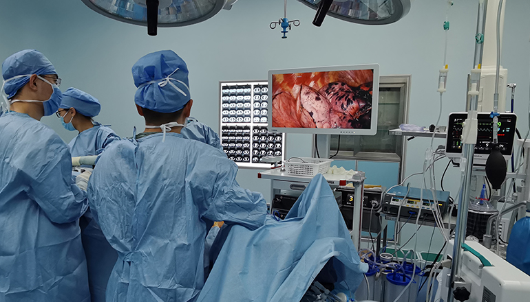
Thoracoscopy, also known as video-assisted thoracic surgery (VATS), is a minimally invasive surgical technique that allows visualization and access to the chest cavity through small incisions. The development of thoracoscopy has revolutionized the field of thoracic surgery, allowing for improved diagnostic and therapeutic capabilities with reduced morbidity and faster recovery times.
One of the primary applications of thoracoscopy is in the diagnosis of pulmonary and pleural diseases. It allows for direct visualization of the lung and pleural cavity, enabling the identification and sampling of abnormal tissue. Biopsy specimens can be obtained using specialized instruments inserted through the thoracoscope, allowing for accurate diagnosis of lung cancer, interstitial lung disease, and other pulmonary conditions.
Thoracoscopy is also used for the treatment of various conditions affecting the chest, including lung and pleural diseases. For example, VATS lobectomy, a surgical procedure in which a portion of the lung is removed, can be performed through small incisions using thoracoscopy. This technique has been shown to have similar outcomes to open surgery but with less pain, reduced morbidity, and shorter hospital stays.
Another application of thoracoscopy is in the management of spontaneous pneumothorax, a condition in which air accumulates in the pleural space causing lung collapse. Thoracoscopy can be used to identify the site of air leak and perform pleurodesis, a procedure that seals the pleural space to prevent further air leaks.

Thoracoscopy can also be used in the management of mediastinal tumors, which are tumors located in the area between the lungs. VATS mediastinal lymph node dissection can be performed using thoracoscopy, allowing for accurate staging and treatment of lung cancer and other mediastinal tumors.
In addition to these clinical applications, thoracoscopy has also been used in the research setting to study the pathophysiology of various lung and pleural diseases. Researchers can use thoracoscopy to directly visualize and sample lung tissue in vivo, allowing for more accurate assessment of disease progression and response to therapy.
Overall, the concept and application of thoracoscopy have greatly improved the field of thoracic surgery, allowing for more precise diagnostic and therapeutic capabilities with reduced morbidity and faster recovery times. This minimally invasive technique has expanded the range of procedures that can be performed in the outpatient setting, enabling patients to receive appropriate care with minimal disruption to their daily lives. With ongoing advancements in technology and technique, it is likely that the applications of thoracoscopy will continue to expand, further improving the management of thoracic diseases.
Leave a Comments