- Shanghai, China
- [email protected]
- +86-21-58189111
looking glass
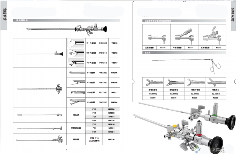
It is the optical part of the cystoscope, which consists of multiple sets of magnifying glasses such as objective lens, intermediate lens, eyepiece and prism.
The objective lens is a plano-convex lens. The magnification of the objective lens and the diameter of the speculum are the keys to determine the size of the inner field of view. If the magnification of the endoscope and the diameter of the endoscope are increased, the internal field of view must also increase.
In the early years, the intermediate mirror was a direct speculum. The structure was simple. There was only one intermediate mirror. The light reflected by the object needed to pass through a long tube diameter to reach the intermediate mirror. Most of the light was absorbed by the tube wall, so the image of the object was blurred. . In order to improve the shortcomings, modern cystoscopes have been composed of most complex lenses (between the objective lens and the intermediate lens), so as to minimize the disappearance of luminosity, and pay attention to correcting chromatic aberration in the production, and strive to achieve a realistic image and color. normal purpose.
The eyepiece is also a plano-convex lens, so that after the object image passes through the above-mentioned groups of lenses, a reduced and upright image is formed before the eyepiece. A lens must be installed at the eyepiece for proper magnification to make the image clearer. The magnification of this lens is closely related to the disappearance of the amount of light. The larger the magnification, the more obvious the disappearance of the light quantity. Generally, it is advisable to magnify 10-20 times. Each type of cystoscope has its own certain magnification.
Triangular prism The triangular prism is used in the speculum, which fundamentally changes the disadvantages of large blind area and small field of view of direct cystoscopy. The prism used in the mirror is a right-angled triangular prism, and the two short sides of the right angle are connected to the objective lens on one side, and the other side is perpendicular to the mirror axis. The object image enters from a short facet and is refracted at a 90-degree angle through the long bevel facet and enters the speculum. Through the intermediate mirror, the eyepiece is reflected into the field of view, but the image of the object seen is upside-down and left-right reversed with the original object. Due to the continuous improvement of modern optical technology, various cystoscopes used clinically are equipped with an Amici prism before the eyepiece.
In order to eliminate the blind spot during cystoscopy to the greatest extent, so as to have a comprehensive and correct view of the interior of the bladder, many authors have continuously improved on the basis of the direct original endoscope, and successively designed and made various reflection angles. Different scopes, such as a 90-degree front sight, a 115-degree front sight, and a 25-degree reverse sight. The operator can choose a speculum with an appropriate reflection angle according to the needs of the examination.

Ureteral intubation and surgical speculum
The optical structure principle of the intubation and surgical speculum is exactly the same as that of the inspection speculum, but the field of view is smaller than that of the inspection speculum. The front end of the intubation speculum is equipped with a diverter, which can be freely raised and lowered by the controller installed at the rear end: according to the position of the ureteral orifice and the lesion, the direction of the ureteral catheter or surgical instruments can be changed at will. The rear end of the speculum is fitted with three small metal tubes. The two smaller holes on the left and right are used for ureteral intubation, and the larger hole in the middle can be used to insert various intravesical surgical instruments (such as electrocautery strips, foreign body forceps, biopsy forceps and scissors, etc.). There is a movable partition attached to the endoscope for intubation, which can be installed during intubation inspection to prevent the left and right ureteral catheters from being bent alternately. However, during surgery, it should be removed to expand the cavity to accommodate various surgical instruments.
Obturator
It is used for inserting the mirror sheath and closing the window of the mirror sheath, so that the cystoscope can be easily introduced or pulled out without damaging the urethral mucosa. The front end of the obturator usually has a small hole or a small groove, so that after the cystoscope is introduced into the bladder, urine will overflow from the small hole or small groove, and the operator can use this to know whether the cystoscope has entered the bladder.
Accessory device
Various types of cystoscopes have certain special accessories. Now only the accessories inherent in general cystoscopy are described as follows:
Power wiring and plug switch: usually a battery or power transformer can be used to connect to the cystoscope through the power wiring. There are two types of latches: latch type and rotary type. Due to the different types of cystoscopes, the above two types of latches cannot be used interchangeably.
Rubber caps: there are big and small; some have holes, some have no holes, they can be put on the small metal tube at the back end of the intubation speculum as needed to prevent water leakage.
Flushing device: German-made Wolf cystoscope, using an automatic spring obturator installed at the rear end of the scope sheath, which automatically closes after pulling out the speculum to prevent fluid from flowing out. A metal flusher needs to be inserted to fill or discharge water. The inch cystoscope flusher is installed on both sides of the rear end of the mirror sheath and is connected by a three-way switch. It can be flushed or discharged as needed during peeping, which is more convenient than the Wolf cystoscope.
Cotton circle: The front end is threaded and can be rolled with cotton for cleaning or drying the cystoscope after use.
Cold light source: It is a bromine light source tungsten light box with brightness adjustment. After the AC power supply, the bromine tungsten lamp can emit a very bright light source.
Light guide: It is composed of extremely fine countless optical fibers, which are used to connect the cold light source and the cystoscope, and transmit the extremely bright light to the front end of the cystoscope. It is an ideal lighting device in the modern cystoscope.
Leave a Comments